
Peripheral osteoma of the maxillary sinus. Case report
DOI:
https://doi.org/10.18004/mem.iics/1812-9528/2026.e24132602Keywords:
Osteoma, Maxillary sinus, Benign neoplasms, Oral surgeryAbstract
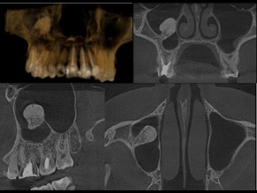
Osteomas are benign tumors characterized by slow growth and composed of compact or cancellous bone. They most commonly occur in the craniofacial region and are uncommon in the paranasal sinuses. They are generally asymptomatic and are usually diagnosed as incidental radiographic findings. Their etiology is not clearly established; however, traumatic, infectious, and genetic factors have been proposed. Diagnosis is confirmed through imaging studies and histopathological analysis, and the most widely accepted treatment is surgical resection when aesthetic or functional alterations are present or when there is a risk of complications. The aim of this study is to describe a clinical case of a peripheral osteoma of the maxillary sinus and its clinical–surgical management. We report the case of a 27-year-old female patient in whom an osseous mass in the maxillary sinus was incidentally detected during an orthodontic evaluation. Computed tomography revealed a well-defined lesion, and surgical resection was performed using a Caldwell–Luc approach under general anesthesia. Complete excision was achieved without complications, with satisfactory postoperative evolution. Histopathological examination confirmed the diagnosis of osteoma, showing mature bone tissue with cortical and trabecular architecture. This case highlights the importance of early radiographic detection and the use of computed tomography for surgical planning. Early resection is a safe procedure that prevents complications and allows exclusion of associated entities such as Gardner syndrome.
Downloads
References
1. Peñaherrera KP, Carlosama FEQ, Bustamante AG. Osteoma de hueso compacto en maxilar superior. Odontología. 2022; 24(2): 24-9. https://dialnet.unirioja.es/servlet/articulo?codigo=8578564
2. Alister JP, Cartes B, Uribe F, Carmine E, Almeida A, Olate S. Resection of fronto-naso-maxillary osteoma: anatomic challenge assisted with stereolithography and piezoelectric system. Int J Morphol. 2017; 35(2): 745-50. http://www.scielo.cl/scielo.php?script=sci_arttext&pid=S0717-95022017000200057
3. Kalfarentzos E, Arapi I, Triantafyllou V, Tosios KI, Christopoulos P. An unusual giant osteoma of the maxillary sinus: case report. SN Compr Clin Med. 2020; 2: 685-8. doi: 10.1007/s42399-020-00278-0
4. Taketomi T, Imayama K, Nakamura K, Kusukawa J. An isolated laminar osteoma arising in the maxillary sinus. Am J Case Rep. 2023; 24: e938904. doi: 10.12659/AJCR.938904
5. Suassuna TM, Abreu NMR, do Nascimento VL, Aguiar ÁP, Sampaio FC, Júnior JWNR. Osteoma extenso em seio maxilar: relato de caso. Arch Health Investig. 2020; 9(4). https://www.archhealthinvestigation.com.br/ArcHI/article/view/4703
6. Patiño-Suárez K, Bravo-Escobar GA, Guzmán-Sánchez M, Brito-Vera JP, Figueroa-Ángel V. Osteoma de senos paranasales con invasión orbitaria: manejo multidisciplinario. An Otorrinolaringol Mex. 2018; 63(4): 173. http://revisionporpares.com/index.php/AORL/issue/download/194/pdf_10
7. Coimbra IMA, Durães RT, Leite LC. Osteoma de seio maxilar invadindo órbita: relato de caso. Braz J Health Rev. 2024; 7(3): e69657. https://ojs.brazilianjournals.com.br/ojs/index.php/BJHR/article/view/69657
8. Licéaga-Escalera C, Del Bosque-Méndez J, Aldape-Barrios B, Montoya-Pérez L, Morelos-Patiño E, González-Olmedo V. Osteoma gigante en mandíbula: reporte de caso. Rev Estomatol Herediana. 2013; 23(4): 206-9. https://www.redalyc.org/pdf/4215/421539379006.pdf
9. Arslan HH, Tasli H, Cebeci S, Gerek M. The management of the paranasal sinus osteomas. J Craniofac Surg. 2017; 28(3): 741-5. doi: 10.1097/SCS.0000000000003397
10. Espinosa Fernández J, Rodríguez Luna R, Ortiz Cruz AB, Amezquita Solórzano JA. Osteoma mandibular periférico. Rev Mex Cir Bucal Maxilofac. 2017; 13(2): 60–4. https://www.medigraphic.com/pdfs/cirugiabucal/cb-2017/cb172e.pdf
11. Durão AR, Chilvarquer I, Hayek JE, Provenzano M, Kendall MR. Osteoma of the zygomatic arch and mandible: report of two cases. Rev Port Estomatol Med Dent Cir Maxilofac. 2012; 53(2): 103–7. 10.1016/j.rpemd.2012.02.006
12. Gondo RM, Costa MDMA, Batista JD, Melo PEC, Silva CM. Osteoma periférico em corpo mandibular: relato de caso. Rev Cir Traumatol Buco-Maxilo-Fac. 2008; 8(1): 23–8. https://pesquisa.bvsalud.org/portal/resource/pt/biblio-873478
13. Irimia IM, Gata A, Puscas IM, Covaliu BF, Albu S. Endoscopic excision of rare large maxillary sinus osteoma: a case report and literature review. Surgeries. 2023; 4: 317-24. doi: 10.3390/surgeries4030032
14. Mafra RP, Vasconcelos RG, Queiroz LMG, Vasconcelos MG. Osteoma maxilar: relato de caso. Rev Bras Cienc Saude. 2014; 18(1): 49–54. doi: 10.4034/RBCS.2014.18.01.07. https://periodicos.ufpb.br/index.php/rbcs/article/view/15250
15. Shinas S, Siriani F, Zisis V, et al. Peripheral osteoma of the maxillary sinus in a patient planned for sinus augmentation. Cureus. 2025; 17(9): e93482. doi: 10.7759/cureus.93482
16. Prabhuji M, Kishore H, Sethna G, Moghe A. Peripheral osteoma of the hard palate. J Indian Soc Periodontol. 2012; 16: 134-7. https://pubmed.ncbi.nlm.nih.gov/22628981/
17. Ahn SY, Ahn CH, Cho KS. Giant osteoma of the mandible causing breathing problem. Korean J Oral Maxillofac Radiol. 2006; 36: 217–20. https://koreascience.kr/article/JAKO200606142043314.pdf
18. Boros LF, Carneiro MIS, Boros LH, Boros LF, Boros PAS. Osteoma compacto central de mandíbula: relato de caso clínico. Odontol Clín-Cient. 2011; 10(1): 89–93. http://revodonto.bvsalud.org/scielo.php?script=sci_arttext&pid=S1677-38882011000100017
19. Martorelli SB de F, Cavalcanti PHAH, Albuquerque R, Guerra EC, Marinho EVS, Martorelli FO. Osteoma exofítico de ângulo mandibular: relato de caso clínico. Odontologia Clín-Científ Recife. 2007; 6(2): 183–6. https://pesquisa.bvsalud.org/portal/resource/pt/lil-509923
Published
Issue
Section
License
Copyright (c) 2026 Memorias del Instituto de Investigaciones en Ciencias de la Salud

This work is licensed under a Creative Commons Attribution 4.0 International License.